
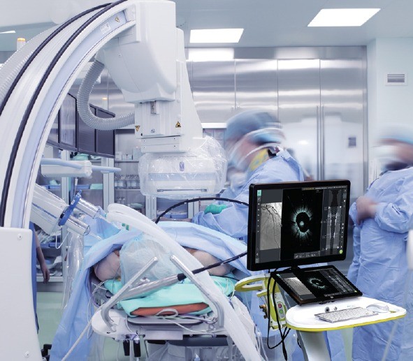
Cardiology guidelines rarely move on the strength of a single trial or a single year of data. The bar for changing how a global field treats coronary artery disease sits deliberately high. Intravascular imaging in percutaneous coronary intervention (PCI) sat in that holding pattern for years, supported by promising data without consensus.
The stance has shifted decisively over the last several years across major societies on both sides of the Atlantic. A decade of randomized evidence, capped by landmark trials between 2023 and 2024, gave societies the grounding to act. The result is a clearer mandate for image-guided PCI, particularly in patients presenting with complex coronary anatomy.
What Changed in the Intravascular Imaging Guidelines?
Both major societies now formally support intravascular imaging during PCI, with stronger language reserved for complex coronary lesions. The 2025 ACC/AHA/SCAI coronary revascularization guideline assigns a Class 1a recommendation for IVI guidance during complex coronary stenting procedures. The 2024 ESC chronic coronary syndromes guideline reinforces the use of image guidance as a reasonable and increasingly expected standard of care.
The Clinical Evidence Behind the Intravascular Imaging Shift
The case for intravascular imaging built slowly across more than a decade of randomized and observational research. Early studies hinted that operators using IVUS or OCT achieved better stent expansion than angiography-only-guided peers. The signal grew stronger as trial populations expanded into more complex anatomy and longer follow-up windows.
ILUMIEN IV, published by Ali and colleagues in 2023, provided the field with a large randomized comparison between OCT- and angiography-guided PCI. The trial enrolled patients with diabetes or complex coronary lesions across more than 80 sites globally. Image guidance resulted in larger minimum stent areas and fewer procedural complications, although the primary clinical endpoint at two years showed no statistically significant difference.
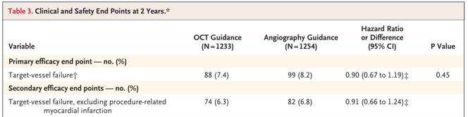
Primary and Secondary Efficacy Endpoints from ILUMIEN IV
Earlier landmark trials had already pointed in the same direction across different lesion subsets and patient populations. Pooled data across thousands of patients consistently favored image-guided PCI on hard endpoints like cardiac death, target-vessel myocardial infarction, and stent thrombosis. Guideline writers finally had the cumulative evidence base they had been waiting for.
The convergence of trial data and real-world registries closed a long-standing question. Image guidance yields durable clinical benefit, particularly in lesion types where procedural stakes are highest.
How U.S. and European Societies Updated Their Recommendations
Two major guideline updates anchor the current global standard for image-guided percutaneous coronary intervention. American and European societies arrived at similar conclusions through parallel review processes that worked from much of the same trial evidence. The convergence matters because operators worldwide now work from compatible recommendations.
The 2024/2025 ACC/AHA/SCAI Guideline for Coronary Artery Revascularization elevated intravascular imaging to a Class 1A recommendation. Coverage extends to selected complex coronary lesions, including left main disease and long-segment cases.
European authorities followed with the 2024 ESC Guidelines for the management of chronic coronary syndromes. The document formalizes intravascular imaging guidelines as part of an optimized PCI strategy in complex coronary anatomy. Specific recommendations address left main intervention, long lesions, and cases in which angiographic ambiguity limits operator decision-making.
Both guidelines stop short of mandating intravascular imaging for every PCI case performed today. Instead, they identify the lesion subsets where evidence is strongest and recommendation language carries the most force. Operators retain clinical judgment for straightforward cases while gaining clear backing for image use in complex ones.
Where Intravascular Imaging Delivers the Strongest Benefit
Guideline endorsement reflects where the evidence sits, not a blanket prescription for every PCI procedure. Certain lesion types consistently benefit from intracoronary imaging more than others. For example, left main coronary artery disease tops the list of high-yield indications. Angiography underestimates left main severity and morphology more often than in other vessels of the coronary tree. Image guidance allows accurate sizing of the main vessel and both branches before any stent commitment.
Bifurcation lesions sit close behind in the hierarchy of clinical benefit. The geometry of side-branch involvement complicates angiographic interpretation.
Long lesions, heavily calcified vessels, and chronic total occlusions round out the high-benefit categories for image guidance. Each presents an anatomy in which pre-PCI assessment changes the treatment strategy in meaningful ways. The Holm bifurcation trial reinforced the broader pattern, showing event reductions that were sustained over two years of follow-up.
Post-stent assessment adds value across nearly every lesion type a cath lab encounters. Confirming expansion, ruling out malapposition, and identifying edge dissections allow operators to address problems before they lead to future readmissions. The clinical payoff compounds as imaging usage becomes routine rather than occasional.
Why Adoption Still Trails the Guidelines
Despite the evidence shift and updated recommendations, intravascular imaging utilization in routine PCI remains low across many regions worldwide. Estimates place global adoption around 20 percent in most reported series. The gap between recommendation and practice has multiple roots worth examining.
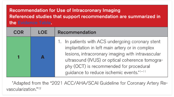
The 2024/2025 ACC/AHA/SCAI Guideline for Coronary Artery Revascularization elevated intravascular imaging to a Class 1A recommendation.
Workflow friction sits near the top of the list of practical barriers. Earlier-generation imaging systems required sterile draping, longer setup times, and more contrast per acquisition. Operators working through high case volumes often defaulted to angiography alone to maintain predictable room turnover.
Catheter size posed its own barrier to broader adoption in complex anatomy. Standard imaging catheters of 2.7F or larger could not cross severely stenosed lesions without prior dilation. The very cases where imaging matters most were sometimes the cases where imaging proved hardest to perform.
Newer technology is actively closing both gaps in ways that match the spirit of recent guidelines. Smaller catheters, faster pullback speeds, and simpler console setup reduce the practical cost of routinely using image guidance. As the equipment becomes easier to use during real procedures, the lag between guideline language and bedside practice narrows steadily.
Align Your Practice With the New Standard
Guideline-supported intravascular imaging is no longer a niche tool reserved for research centers and complex referral cases. The evidence base is settled, the recommendations are explicit, and the technology has matured to fit routine cath lab workflow. What remains is for individual practices to bridge the adoption gap using tools designed to meet the standard now described by the guidelines.
The Gentuity HF-OCT Imaging System and Vis-Rx PRIME Micro-Imaging Catheter were engineered with that gap in mind from the start. Faster pullback, smaller catheter profile, and simpler setup remove the friction that kept earlier-generation systems from broader use. Discover how HF-OCT is designed to meet the standard that guidelines are now setting at Gentuity.com.
Sources
Ali ZA, Landmesser U, Maehara A, et al; ILUMIEN IV Investigators. Optical Coherence Tomography-Guided versus Angiography-Guided PCI. N Engl J Med. 2023;389(16):1466-1476. doi:10.1056/NEJMoa2305861.
Holm NR, Andreasen LN, Neghabat O, et al; OCTOBER Trial Group. OCT or Angiography Guidance for PCI in Complex Bifurcation Lesions. N Engl J Med. 2023;389(16):1477-1487. doi:10.1056/NEJMoa2307770.
Get all the latest updates
Sign up for updates, HF-OCT clinical insights, and company news.
"*" indicates required fields

