

Intravascular optical coherence tomography (OCT) has reshaped how interventional cardiologists assess coronary anatomy before, during, and after percutaneous coronary intervention (PCI). The modality uses near-infrared light to generate cross-sectional images with axial resolution of approximately 10 microns, far exceeding that of angiography or intravascular ultrasound. Clinical guidelines increasingly recommend image-guided PCI for complex lesions, yet adoption has lagged behind the supporting evidence.
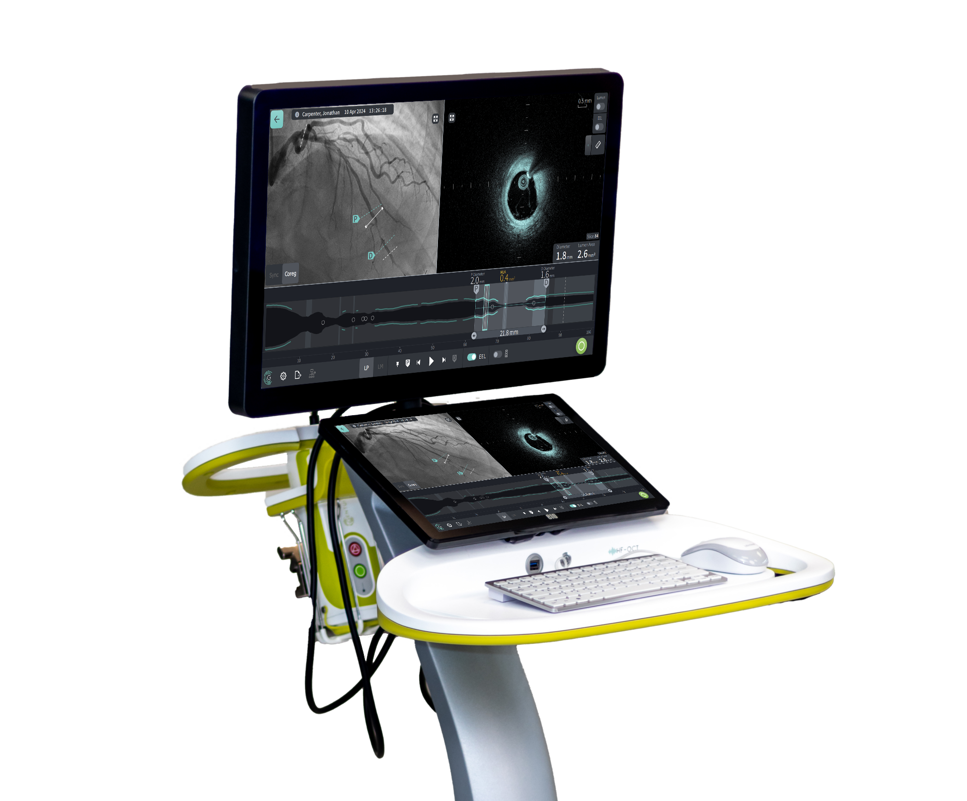
High-frequency OCT, or HF-OCT, by Gentuity, represents the next-generation evolution of the platform. Engineered around a 1.8F micro-imaging catheter and a one-second pullback, the system aims to address limitations that have constrained pre-PCI imaging in tight or tortuous vessels. Clinicians evaluating coronary disease can now visualize the full artery in a single, rapid acquisition before making any treatment decisions.
What Is High-Frequency Optical Coherence Tomography?
HF-OCT is a coronary imaging technology that pairs a swept-source laser operating near 1310 nm with an ultra-low-profile catheter to capture up to 100 mm of vessel in one second. The catheter measures 1.8F (0.6 mm), enabling crossing of severely stenosed lesions without pre-dilation. The expanded field of view exceeds 14 mm in diameter, supporting complete imaging of left main and ostial segments that other OCT systems cannot fully capture.
How HF-OCT Builds on Standard Optical Coherence Tomography
Optical coherence tomography entered the cath lab in the late 2000s as a high-resolution way to visualize coronary anatomy from within. Resolution that sharp lets clinicians identify plaque type, vessel size, and stent apposition with a clarity that angiography cannot match.
Standard OCT, however, was built around a 2.7F imaging catheter and a pullback of about two seconds. The catheter size limits the crossing of tight lesions before any vessel preparation. Pullback length tops out at around 75 mm, which often requires a second acquisition for longer segments.
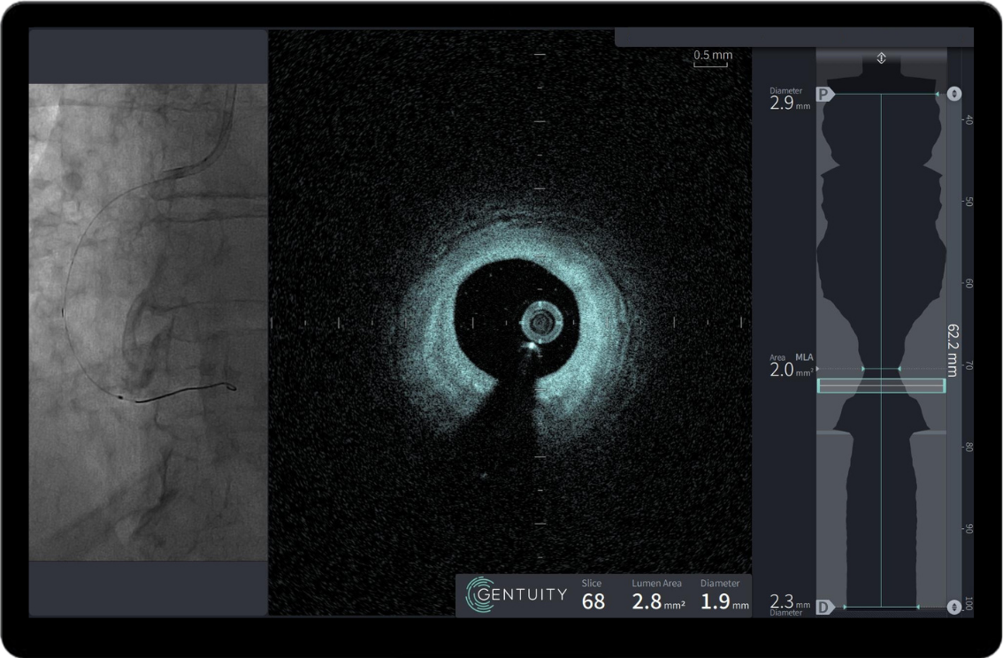
Saline-only HF-OCT Imaging of the right coronary artery
HF-OCT was developed by members of the original LightLab OCT team to address those gaps directly. The catheter is 1.8F, roughly half the cross-sectional area of legacy systems. Pullback speed climbs to 100 mm per second, covering the complete vessel in a single second.
The first-in-human study by Hiram Bezerra, MD and colleagues showed HF-OCT could capture clear images in severely stenosed arteries without balloon preparation. Faster acquisition also reduces the contrast burden tied to each pullback. Optical coherence tomography, in its high-frequency form, becomes a tool clinicians can reach for earlier in the procedure rather than only after intervention.
What Makes HF-OCT Different in the Cath Lab
Several engineering choices set HF-OCT apart from earlier OCT platforms. The differences show up in how the catheter handles tight anatomy, how quickly images appear, and how the system fits into routine workflow. Each contributes to a more practical pre-PCI imaging experience.
Ultra-low-profile catheter: The Vis-Rx PRIME catheter measures 1.8F, allowing it to cross severely narrowed lesions that other imaging catheters cannot reach.
One-second pullback: A full 100 mm of coronary artery is captured in a single second, well beyond the 75 mm typical of standard OCT.
Reduced contrast burden: Faster acquisition supports imaging runs with as little as 5 mL of contrast, or saline alone in select cases.
Plug-and-play setup: The probe interface module mounts to the bedrail and requires no sterile bags enabling a very simple setup.
AI-assisted measurements: Automated lumen sizing and StentX expansion analysis surface key numbers to support confident treatment decisions.
The combination matters because pre-PCI imaging changes treatment decisions far more often than post-PCI imaging alone. Simple setup, smooth delivery, and rapid imaging make the technology easy to incorporate into workflow.
Where Optical Coherence Tomography Helps Most During PCI
Pre-PCI assessment is where image guidance does its heaviest work. Seeing lesion morphology before modification or stent selection prevents committing to a potential suboptimal strategy and informs device sizing, landing zones, and whether to pursue direct stenting or lesion preparation. Calcium, lipid pools, and thrombus all influence strategy in ways angiography cannot reveal.
Complex lesions raise the stakes further. Heavily calcified vessels, long diffuse disease, bifurcations, and left main anatomy each benefit from a clear picture of the full segment. HF-OCT is especially useful when small vessel size or tight stenosis would preclude the use of conventional catheters.
Post-stent imaging closes the loop. Operators can confirm expansion, check for malapposition, and identify edge dissections that may need additional treatment. Stent‑expansion targets in both US and EU society guidelines consistently point to a key threshold: achieving ≥80% of the reference vessel area is linked with stronger long‑term clinical outcomes.
The multicenter study conducted by Donald Quimby, Jr., et al. found HF-OCT delivered reliable image quality across pre- and post-intervention pullbacks in an all-comers population. Image clarity held up even in arteries with severe stenosis.
What Clinicians Gain From Faster, Smaller Imaging
Speed changes how often imaging gets used during a procedure. A one-second pullback fits into the natural rhythm of PCI rather than interrupting it. Operators reach for the tool more readily when setup and acquisition feel routine instead of cumbersome.
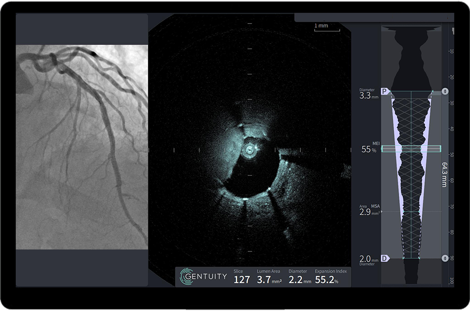
HF-OCT’s StentX software quantifies and visualizes stent expansion, one of the most important determinants of successful stent outcomes.
A smaller catheter size enables cases that previously required pre-dilation or vessel modification to be performed first. As a result, crossing the lesion before any manipulation preserves the true picture of the disease. Treatment strategy gets built on what the artery actually looks like, not what it looks like after a balloon has passed.
Contrast reduction matters for patients with renal concerns or for those undergoing long procedures. Lower per-run volumes give operators flexibility to image more often without raising total contrast exposure. Saline-only acquisitions extend that flexibility further in select anatomies.
Workflow simplicity ties the benefits together, supporting broader adoption of optical coherence tomography. When the console rolls in, plugs in, and images in seconds, without cumbersome setup and workflow, the barrier to routine intravascular imaging drops considerably. Faster acquisition also means more pullbacks per case when clinical judgment calls for them.
Bring HF-OCT Into Your PCI Practice
HF-OCT represents a practical step forward for clinicians who want intravascular imaging to work as guidelines suggest. The technology pairs high-resolution detail with the speed and accessibility a busy cath lab actually needs. Adoption hinges on tools that fit the procedure rather than slow it down.
The Gentuity HF-OCT Imaging System and Vis-Rx PRIME Micro-Imaging Catheter deliver that combination today. Clinical evidence and global guideline support continues to grow across pre-PCI assessment, complex lesion guidance, and post-stent optimization. Learn more about the Gentuity HF-OCT Imaging System at Gentuity.com
Sources
Bezerra HG, Quimby DL Jr, Matar F, Mohanty BD, Bassily E, Ughi GJ. High-frequency optical coherence tomography (HF-OCT) for preintervention coronary imaging: a first-in-human study. JACC Cardiovasc Imaging. 2023;16(7):982-984. doi:10.1016/j.jcmg.2023.01.013.
Quimby DL Jr, Rothstein ES, Richmond HCT, et al. Efficacy and Safety of High-Frequency Optical Coherence Tomography (HF-OCT) for Coronary Imaging: A Multicenter Study. J Soc Cardiovasc Angiogr Interv. 2025;4(3 Part A):102577. doi:10.1016/j.jscai.2025.102577.
Get all the latest updates
Sign up for updates, HF-OCT clinical insights, and company news.
"*" indicates required fields

